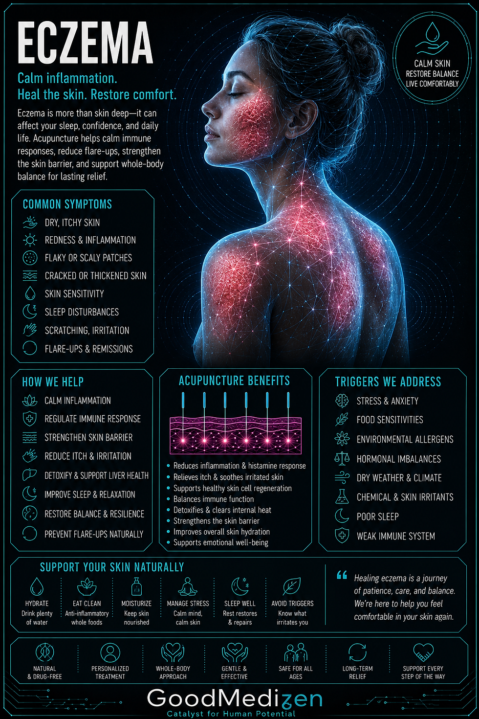
Your Skin Is Trying to Tell You Something
Eczema is one of those conditions where the treatment focus almost always ends up in the wrong place. The itching, the rash, the inflamed patches — those are real and they're miserable. They're the message, not the problem. Skin doesn't become chronically inflamed for no reason. Something is driving that inflammation, and until we find it, the best you can hope for is managing flares rather than actually resolving them.
Most people with eczema have been through the steroid cream cycle. It works for a while, then stops working, or you need increasingly stronger versions, or it works on one patch while another one appears somewhere else. That's not a failure of the medication — it's the predictable result of suppressing a symptom without addressing what's causing it.
What Eczema Actually Is
Eczema — clinically called atopic dermatitis — is a chronic inflammatory skin condition characterized by a defective epidermal barrier, immune dysregulation, and a heightened inflammatory response to triggers that wouldn't affect uncompromised skin.
The word "atopic" refers to a hereditary tendency toward allergic conditions — eczema, asthma, and allergic rhinitis often cluster in the same families and in the same person, because they share an underlying immune pattern. That immune pattern, called a Th2-dominant response, means the immune system preferentially launches allergic-type reactions rather than the pathogen-clearing Th1 response. This isn't a malfunction — it's a regulatory bias that's been tipped in one direction.
The skin barrier defect is central to the eczema story. In most people with atopic dermatitis, mutations or dysfunction in a protein called filaggrin compromise the integrity of the skin's outer layer. Filaggrin is responsible for binding keratinocytes (skin cells) together and maintaining the lipid matrix that keeps moisture in and irritants out. When filaggrin is deficient or dysfunctional, the skin becomes permeable — moisture escapes, allergens enter, and the immune cells just beneath the surface stay in a constant low-grade state of activation.
When an irritant or allergen penetrates the compromised barrier, those primed immune cells release inflammatory signaling molecules — IL-4, IL-13, IL-31, and TSLP (thymic stromal lymphopoietin). IL-31 is the one most directly responsible for the itch signal — it binds to receptors on sensory nerves and triggers the itch-scratch cycle. Scratching further damages the barrier, creating more entry points for irritants, perpetuating the inflammation. The cycle becomes self-reinforcing.
The Roots We Actually Find
Gut dysbiosis and the gut-skin axis. The gut microbiome has a profound influence on immune regulation — particularly the balance between Th1 and Th2 responses. Dysbiosis and intestinal permeability allow bacterial fragments (LPS endotoxins) to enter systemic circulation, driving the kind of low-grade immune activation that primes skin toward inflammatory responses. Studies have found significantly altered gut microbiome composition in people with eczema, often with reduced Lactobacillus and Bifidobacterium species that support regulatory immune function. Gut healing is almost always part of eczema treatment that holds long term.
Food sensitivities and IgG-mediated reactions. IgE-mediated food allergies (immediate, anaphylactic-type) are obvious — but delayed IgG-mediated food sensitivities are subtler and far more commonly linked to eczema flares. These reactions can occur hours to days after exposure, making them difficult to identify without testing. Common culprits: dairy, eggs, wheat, soy, tree nuts, and citrus — though individual patterns vary significantly.
Histamine dysregulation. Histamine is an inflammatory signaling molecule involved in both allergic responses and gut motility. In some people, either excessive histamine production or impaired histamine clearance (due to low levels of the enzyme DAO — diamine oxidase, produced in the gut lining) creates a chronic histamine excess. High-histamine foods (fermented foods, aged cheeses, alcohol, certain fish) can exacerbate inflammatory skin reactions in this pattern.
Nervous system dysregulation. The skin and nervous system are intimately connected — they develop from the same embryological tissue layer (ectoderm). Chronic stress, sympathetic nervous system dominance, and impaired vagal tone all amplify inflammatory signaling in skin. Eczema is notoriously stress-reactive, and the mechanism isn't just behavioral — stress hormones directly modulate the inflammatory cytokines driving the eczema pattern.
Nutrient deficiencies. Vitamin D modulates the Th1/Th2 balance and supports skin barrier function — deficiency is consistently associated with more severe eczema. Zinc is required for skin repair and immune regulation. Omega-3 fatty acids are incorporated into skin cell membranes and shift the inflammatory balance toward resolution.
Where TCM Comes In
Chinese medicine has recognized the internal origins of skin conditions for centuries. The skin in TCM is considered the outer expression of what's happening in the internal organs — particularly the Lung (which governs the skin and the wei qi, or defensive energy at the body's surface), the Spleen (which governs transformation and movement of fluids), and the Liver (which governs the smooth flow of qi and blood).
Wind-Heat or Damp-Heat in the Skin. Acute flares with significant redness, oozing, intense itch. Corresponds to histamine-driven reactions with active immune activation and elevated inflammatory cytokines.
Spleen Qi Deficiency with Dampness. Chronic eczema with pale, doughy, or weeping lesions, often accompanied by digestive symptoms — bloating, loose stools, fatigue. Corresponds to the gut dysbiosis and impaired barrier function pattern.
Blood Deficiency and Wind. Chronic, dry, thickened eczema — lichenified patches, intense itch that's worse at night. Corresponds to impaired skin barrier, chronic inflammation with inadequate tissue nourishment, and elevated IL-31 driving the night-time itch signal.
Liver Qi Stagnation with Heat. Eczema that clearly worsens with stress and emotional pressure, often with irritability, tension headaches, and disrupted sleep. Corresponds to the cortisol-driven inflammatory amplification pattern.
How We Approach It
Acupuncture reduces systemic inflammation, regulates the HPA axis (stress response system), and calms the immune hypersensitivity driving the Th2-dominant pattern. Specific point protocols address the TCM pattern driving the presentation, while others directly modulate the itch signal through effects on sensory nerve transmission.
Chinese herbal medicine is particularly powerful for eczema. Classical formulas for damp-heat skin conditions have been used for centuries with strong clinical results, and several have been studied in RCTs. Formulas are always tailored to the individual pattern.
Functional medicine identifies the specific drivers. We look at food sensitivities (IgG panel), comprehensive stool analysis for gut dysbiosis and permeability markers, vitamin D levels, and nutrient status. We build a targeted plan based on what we find.
Topical support — beyond standard pharmaceutical emollients, we can recommend specific barrier repair approaches with research support: colloidal oatmeal, calendula, and ceramide-containing formulations.
When to Consider Us
- Your eczema cycles on and off and you want to break the cycle rather than manage it indefinitely
- Topical steroids have stopped working or you don't want to keep using them
- Your eczema clearly worsens with stress, diet, or seasonal changes
- You have other atopic conditions alongside the eczema — asthma, hay fever, food allergies
- Your child has eczema and you want a non-pharmaceutical approach
- You've noticed gut symptoms, food sensitivities, or frequent illness alongside the skin issues
Selected References
- Lee, S. Y., et al. (2016). Microbiome in the gut-skin axis in atopic dermatitis. Allergy Asthma Immunol Res, 10(4), 354–362.
- Lai, R., et al. (2021). Acupuncture for atopic eczema: A systematic review and meta-analysis. ECAM, 2021.
- Weidinger, S., & Novak, N. (2016). Atopic dermatitis. Lancet, 387(10023), 1109–1122.
- Kim, J. E., & Kim, H. S. (2019). Microbiome of the skin and gut in atopic dermatitis. Ann Dermatol, 31(5), 489–496.